The pituitary gland is within the sella turcica or the hypophyseal fossa. This structure is present near the center at the base of the cranium and is fibro-osseous. The anatomical boundaries of the gland have clinical and surgical significance. Sella turcica is a concave indentation in the sphenoid bone.
What gland is located in the sella turcica of the sphenoid bone?
Your pituitary (hypophysis) is a pea-sized endocrine gland at the base of your brain, behind the bridge of your nose and directly below your hypothalamus. It sits in an indent in the sphenoid bone called the sella turcica.
Which gland is located in the sella turcica quizlet?
Located at the base of the brain, the pituitary gland is protected by a bony structure called the sella turcica of the sphenoid bone.
What organ is in the sella turcica?
The sella turcica is a midline depression in the sphenoid bone which contains the pituitary gland and distal portion of the pituitary stalk.What is the sella turcica quizlet?
sella turcica. depression in the sphenoid bone where the pituitary gland is located. hypophyseal fossa. The Hypophyseal Fossa is “seat of the saddle” (Sella Turcica) part of the Sphenoid bone. It is the groove that is the deepest part of the body and it holds the Pituitary gland.
What is the sella turcica and its significance?
During embryological development, the sella turcica area is the key point for the migration of the neural crest cells to the frontonasal and maxillary developmental fields. The neural crest cells are involved in the formation and development of sella turcica and teeth.
Where is sphenoid bone?
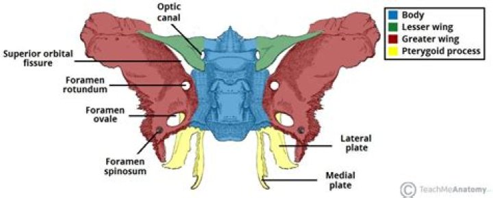
The sphenoid is an unpaired bone. It sits anteriorly in the cranium, and contributes to the middle cranial fossa, the lateral wall of the skull, and the floor and sides of both orbits. It has articulations with twelve other bones: Unpaired bones – Occipital, vomer, ethmoid and frontal bones.
Which endocrine gland is housed in the sella turcica of the sphenoid bone quizlet?
The pituitary gland lies in the sella turcica of the sphenoid bone— a bony cavity at the base of the brain.Why is the sella turcica important?
The sella turcica serves as an important anatomical reference in orthodontics partly because the s-point, placed centrally in the sella region, is a central fix point in cephalometric analysis and partly because the contour of the anterior wall is used in evaluation of craniofacial growth.
What endocrine organ is found in the roof of the third ventricle?Anatomy of the pineal gland The pineal gland develops from the roof of the diencephalon, a section of the brain, and is located behind the third cerebral ventricle in the brain midline (between the two cerebral hemispheres).
Article first time published onWhich gland is posterior to the sternum?
The thymus is a soft organ with two lobes that is located anterior to the ascending aorta and posterior to the sternum.
What structure is found within the sella turcica depression?
The rounded depression in the floor of the sella turcica is the hypophyseal (pituitary) fossa, which houses the pea-sized pituitary (hypophyseal) gland. The greater wings of the sphenoid bone extend laterally to either side away from the sella turcica, where they form the anterior floor of the middle cranial fossa.
What bone contains the depression called the sella turcica what is located in the Depression?
The Pituitary Gland The sphenoid bone lies at the base of your skull, and in this bone is a small, cup-shaped depression called the sella turcica (“Turkish saddle”). Lying in this depression is a round ball of tissue, about 1.3 cm (0.5 in) in diameter, called the pituitary gland or hypophysis (Figure 1.9).
Which feature is found only on thoracic vertebrae?
The characteristic feature of thoracic vertebrae is the presence of joints that articulate with ribs. A mid-thoracic vertebra (shown here) has two joint facets on the vertebral body for the heads of adjacent ribs, and a third joint facet on the transverse process for the neck of a rib.
What attaches to the sphenoid bone?
On the base of the sphenoid bone, several muscles attach to it’s legs, the medial and lateral pterygoid processes. As the name suggests, the pterygoid muscles, important for chewing and mastication, attach here.
What is in the sphenoid bone?
a median portion, known as the body of sphenoid bone, containing the sella turcica, which houses the pituitary gland as well as the paired paranasal sinuses, the sphenoidal sinuses. two greater wings on the lateral side of the body and two lesser wings from the anterior side.
What is sphenoid bone in anatomy?
The sphenoid is just one of the twenty-two bones that form the skull and essentially helps to connect the neurocranium to the facial skeleton. It is a single bone in the midline of the cranial cavity situated posterior to the frontal bone but anterior to the occipital.
Which endocrine gland stores and releases oxytocin and antidiuretic hormone?
The pituitary gland is divided into two distinct structures with different embryonic origins. The posterior lobe houses the axon terminals of hypothalamic neurons. It stores and releases into the bloodstream two hypothalamic hormones: oxytocin and antidiuretic hormone (ADH).
What is the target organ of TSH?
Endocrine gland/ source of hormoneHormoneTarget organ or tissueAnterior pituitary (adenohypophysis)LH (luteinizing hormone)Ovaries / testes (Leydig cells)GH (growth hormone)All tissuesTSH (thyroid stimulating hormone)Thyroid glandProlactinMammary gland
Which system only Innervates one organ and a limited number of cells within that organ?
An efferent nerve fiber innervates only one organ and a limited # of cells within that organ, whereas hormones circulate throughout the body and can have widespread effects.
Which of the following glands is located on the third ventricle of the brain?
The pineal gland, also called pineal body or epiphysis cerebri, is a small cone-shaped structure that extends posteriorly from the third ventricle of the brain.
Is the pineal gland an organ?
PINEAL PHYSIOLOGY The pineal gland in humans is a small (100-150mg), highly vascularized, and a secretory neuroendocrine organ. It is located in the mid-line of the brain, outside the blood-brain barrier and attached to the roof of the third ventricle by a short stalk.
What endocrine organ is located in the anterior neck?
The thyroid gland and parathyroid glands are located in front of the neck, below the larynx (voice box). The thyroid plays an important role in the body’s metabolism.
Which gland is divided into anterior and posterior lobes quizlet?
The pituitary gland is divided into two distinct lobes: the anterior (adenohypophysis) and the posterior (neurohypophysis).
Which gland is divided into anterior and posterior?
The pituitary gland can be divided into two different parts: the anterior and posterior lobes.
Where is the thymus gland situated?
The thymus gland is in the chest, between the lungs and behind the breastbone (sternum). It is just in front of, and above, the heart. The thymus makes white blood cells called T lymphocytes (also called T cells). These are an important part of the body’s immune system, which helps us to fight infection.
Which thoracic vertebrae contain costal facets on the transverse process?
The tenth thoracic vertebra (T-10) usually has a complete, superiorly placed costal facet on each side of the vertebral body and costal articulations on the transverse processes.
Which of these features is found in lumbar vertebrae?
Distinguishing features of the lumbar vertebrae include a thick and stout vertebral body, a blunt, quadrilateral spinous process for the attachment of strong lumbar muscles, and articular processes that are oriented differently than those found on the other vertebrae.
What feature is found in a lumbar vertebra?
The lumbar vertebrae (L1 to L5) have fairly long transverse processes and large, flat, rectangular-shaped spines (see Figure 3.11C). The main distinguishing feature of the lumbar vertebrae is the orientation of the facets on the superior and inferior articular processes.