Abstract. Image processing or digital image manipulation is one of the greatest advantages of digital radiography (DR). Preprocessing depends on the modality and corrects for system irregularities such as differential light detection efficiency, dead pixels, or dark noise.
What is the purpose of image processing operations in digital radiography systems?
a wide histogram demonstrates ___________ contrast, whereas a narrow histogram shows ________ contrast. contains stored data to substitute new values for each pixel during processing. The proper LUT will provide the proper grayscale, regardless of variations in kvp and mAs, resulting in consistent images.
Why is it important to use the correct processing parameters when processing a digital image?
One of the advantages of digital processing is that the processing parameters (factors) can be selected to produce images with different contrast characteristics. … Like the characteristic curve of film, the slope of the curve at every point represents how the contrast will be changed by the processing.
What is digital processing in radiography?
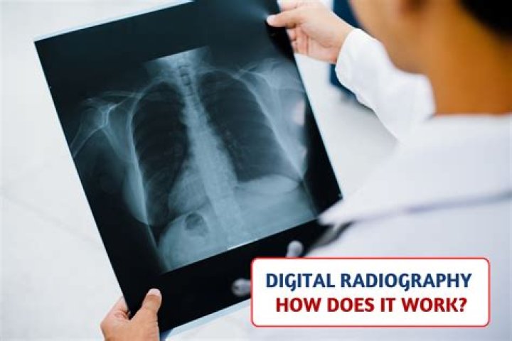
Digital radiography (DR) is an advanced form of x-ray inspection which produces a digital radiographic image instantly on a computer. This technique uses x-ray sensitive plates to capture data during object examination, which is immediately transferred to a computer without the use of an intermediate cassette.What is the purpose of automatic rescaling?
FIGURE 7-2 Automatic rescaling is employed during histogram analysis to maintain consistent image brightness despite overexposure or underexposure of the IR.
What are the features of digital image processing?
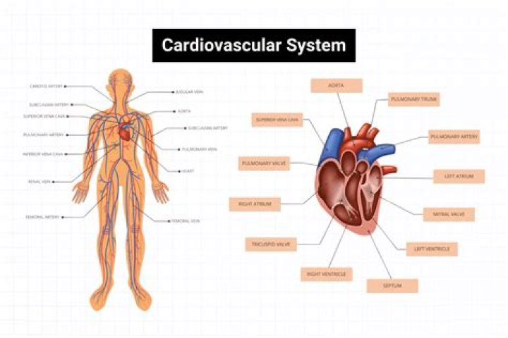
Four Characteristics of a Digital Image A digital image has four basic characteristics or fundamental parameters: matrix, pixels, voxels, and bit depth. A digital image is made up of a 2D array of numbers called a matrix. A matrix is a rectangular array of numbers, symbols, or expressions arranged in rows and columns.
How does digital image processing work?
Digital image processing deals with manipulation of digital images through a digital computer. … The input of that system is a digital image and the system process that image using efficient algorithms, and gives an image as an output. The most common example is Adobe Photoshop.
What are the good characteristics of a digital radiography system?
There are several advantages of digital x-ray imaging over analog film imaging that can benefit the clinician5: reduced time, reduced radiation, ability to take multiple exposures without repositioning the sensor, storage and maintenance of the images, and electronic transmission of images.What are the advantages of digital radiography?
Digital Radiography Advantages: Reducing Cost and Space Reduced radiation. Reduced cost due to the elimination of chemical processors, processor maintenance, and filing and mailing jackets. Reduced space requirement — no dark room is required, and the need to dedicate space for cabinets of analog images is eliminated.
What does digital imaging require?Digital imaging requires an x-ray unit, sensors to capture the images, computer hardware and software to view, store, and transfer images, and if an indirect digital system, a scanning device.
Article first time published onWhich of these processes identifies the values of interest in a radiograph before processing?
Histogram analysis identifies the values of interest in a radiograph before processing.
What is pixel pitch in radiography?
Pixel pitch is the distance between the centers of two consecutive photon-detecting pixels. It is measured in micrometers (µm) and generally ranges between 75 and 200µm. To better understand the importance of pixel pitch, it is necessary to distinguish between detectivity and resolution.
What is automatic rescaling and what is its relationship to exposure factors?
Automatic rescaling means that images are produced, regardless of the amount of exposure, with: uniform size and shape. uniform contrast and density. … provide appropriate brightness and contrast.
What is edge enhancement in radiography?
Edge enhancement is an image processing filter that enhances the edge contrast of an image to improve its apparent sharpness. This feature has the effect of creating subtle bright and dark highlights on either side of any edges in the image, leading the edge to look more defined.
What is dynamic range in radiography?
The dynamic range describes the range of x-ray intensities a detector can differentiate. A high dynamic range provides the discrimination between small differences in x-ray attenuation. A current CT scanner has approximately a dynamic range of 1,000,000 to 1 and 1,100 views or projections a second.
What is preprocessing of image?
Image preprocessing are the steps taken to format images before they are used by model training and inference. This includes, but is not limited to, resizing, orienting, and color corrections. … Thus, a transformation that could be an augmentation in some situations may best be a preprocessing step in others.
What are the types of digital image processing?
Digital image processing techniques are typically classified into three categories. These categories include image generation, enhancement, and restoration. Generation techniques help project and recognize a scanned image, while the process of enhancing an image involves improving contrast, brightness and hue.
What are the principal advantages of digital radiography over conventional radiography?
Advantages over Conventional Radiography Images produced with conventional radiography have limited dynamic range, but digital radiography allows for many exposures to build up a high contrast image. A digital radiograph can be easily copied, e-mailed, and enhanced without degradation.
What are the advantages of digital imaging technology quizlet?
One advantage of a digital imaging system is the superior gray-scale resolution that results. Digital subtraction is an advantage in digital imaging because distracting background information is eliminated from the image. The manipulation of the original digital images can be considered a legal issue.
What are the types of digital radiography?
There are two types of digital imaging systems used in intraoral radiography – computed radiography (CR) and direct radiography (DR). CR uses a photostimulable phosphor (PSP) plate to capture the image.
What is bit depth in digital radiography?
BIT DEPTH is determined by the number of bits used to define each pixel. The greater the bit depth, the greater the number of tones (grayscale or color) that can be represented. Digital images may be produced in black and white (bitonal), grayscale, or color.
How is the latent image formed in digital radiography?
When exposed to x-rays, electrons in the phosphor plate are excited into a higher energy state, forming a latent image. … When the trapped electrons absorb the laser energy, they emit light as they return to their ground state.
How does digital radiography differ from traditional radiography?
With the addition of computer technology, digital radiography has become a much more efficient, cost effective, and an even safer method of producing diagnostic images. … While traditional X-rays are considered safe, digital X-rays produce 80% less radiation than traditional.
What is quantization in radiology?
Quantization, involved in image processing, is a lossy compression technique achieved by compressing a range of values to a single quantum value. When the number of discrete symbols in a given stream is reduced, the stream becomes more compressible.
Are intensifying screens used in digital radiography?
The intensifying screen as part of a film screen system has been an important component in radiology to reduce the radiation dose of the patient. Today, the conventional film cassette is being replaced by an imaging plate used in digital systems. … Mammography cassettes are equipped with single screens.
How often must processing solutions be replenished?
How often should the processing solutions be replenished? Fresh chemicals produce the best radiographs. To maintain freshness, film processing solutions must be replenished daily and changed every 3 to 4 weeks.
What are the three 3 major factors affecting radiographic image quality?
Besides, the above, was should know that contrast of the radiographic image is controlled by three factors viz., (1) film contrast, (2) processing chemicals and (3) radiation factors of objective contrast.
What causes histogram analysis errors?
In computed radiography, histogram analysis errors can occur because of the image being improperly centered to the IR or all collimated borders not shown or aligned accurately.
What is most often the weakest link in the digital imaging chain?
what is the weakest link in the digital imaging chain? the more manipulation of the before being sent to PACS, the greater the loss of information.
What is monitor dot pitch?
The term “dot pitch” refers to the distance between like-colored phosphors on a video monitor. The smaller the dot pitch, the finer the image can be. … Higher values such as 0.35mm are usually found only on older monitors, or on larger displays intended to be seen from a distance.
What is the resolution of an xray?
Radiographic Imaging Digital radiographs generally have a ma- trix size of 2000 × 2500. For a 35 × 43-cm cassette, this digital matrix corresponds to a pixel size of 175 μm and a limiting resolution of three line pairs per millimeter.