The dura is a thin layer of tissue that covers and protects the spinal cord. It lies in between the spine (the bone) and the spinal cord (nerve tissue). Durotomy, which is an incision in the dura, can be a planned part of the surgical technique.
What is the function of the dura?
The dura mater is a sac that envelops the arachnoid and has been modified to serve several functions. The dura mater surrounds and supports the large venous channels (dural sinuses) carrying blood from the brain toward the heart. The dura mater is partitioned into several septa, which support the brain.
What is the dura made of?
The dura mater is made up of fibroblasts and large amounts of extracellular collagen. The dura mater is composed of two layers: the periosteal/endosteal layer and the meningeal layer. The dural venous sinuses are between these two layers.
Is the dura part of the spinal cord?
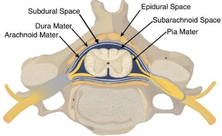
The spinal dura mater is a fibrous, non-adherent, tough layer surrounding the spinal cord. It is separated from the wall of the vertebral canal by the epidural space.Where is the dura located?
The dura mater is the layer that is present on the outermost end of the meninges, situated directly beneath the skull and the bones of the vertebral column.
Is the dura mater pain sensitive?
In this observational study, we confirmed that dura of the skull base and dura of the falx cerebri are sensitive to pain and that their mechanical stimulation induced pain mainly referred in the sensory territories of the V1 and V3 divisions of the trigeminal nerve.
What does dura mean in medical terms?
Dura: The outermost, toughest, and most fibrous of the three membranes (meninges) covering the brain and the spinal cord. Dura is short for dura mater (from the Latin for hard mother). Also called the pachymeninx (singular) or pachymeniges (plural). Epidural means outside the dura.
Where is CSF located?
CSF is secreted by the CPs located within the ventricles of the brain, with the two lateral ventricles being the primary producers. CSF flows throughout the ventricular system unidirectionally in a rostral to caudal manner.Where is your dural sac?
The sleeve of dura is called the dural sac. It’s open at the top end, and closed at the bottom. Here at the base of the skull, the dural sac passes through the foramen magnum, becoming continuous with the layer of dura that surrounds the brain.
Where is CSF produced?CSF is produced mainly by the choroid plexus epithelium and ependymal cells of the ventricles and flows into interconnecting chambers; namely, the cisterns and the subarachnoid spaces.
Article first time published onWhat kind of tissue is the dura?
It is composed of dense fibrous tissue, and its inner surface is covered by flattened cells like those present on the surfaces of the pia mater and arachnoid.
Is the cranial dura the same as the spinal dura?
The dura mater is a membrane that envelops the arachnoid mater. … When it covers the spinal cord it is known as the dural sac or thecal sac. Unlike cranial dura mater, spinal dura mater only has one layer, known as the meningeal layer. The potential space between these two layers is known as the epidural space.
What are the four regions of the spinal dura mater?
The spinal cord and spine are divided into 4 regions from top to bottom: cervical, thoracic, lumbar, and sacral.
What is the center of the brain called?
The cerebrum (front of brain) comprises gray matter (the cerebral cortex) and white matter at its center. The largest part of the brain, the cerebrum initiates and coordinates movement and regulates temperature.
Is dura mater avascular?
Epidural Space and Dura Mater While it was once thought that the dura was avascular, it is actually highly vascular in nature, as the major vessels that supply it run in the epidural space deep to the skull.
What is CSF function?
Cerebrospinal fluid (CSF) is a clear, colorless liquid found in your brain and spinal cord. … CSF helps protect this system by acting like a cushion against sudden impact or injury to the brain or spinal cord. CSF also removes waste products from the brain and helps your central nervous system work properly.
What is the dura mater mater?
(DER-uh MAY-ter) The tough outer layer of tissue that covers and protects the brain and spinal cord and is closest to the skull. The dura mater is one of the three layers that form the meninges.
What is the meaning of the prefix Dura -?
In Latin, dura means “hard or thick.” Definitions of dura. the outermost (and toughest) of the 3 meninges. synonyms: dura mater. type of: meninges, meninx.
Why is dura mater called tough mother?
Definition: dura mater. The outermost layer of three meninges, or membranes, that surround the spinal cord and the brain. In Latin, it means “tough mother.” The name is apt, because the membrane is thick and strong, and normally firmly attached to the inner side of the skull.
What are Dural signs?
Dural signs and symptoms are those that are related to the increase of dural irritation: traction exerted from a distance (straight leg raising and neck flexion) pulls on the inflamed dura or, via the dural ligaments, on the posterior longitudinal ligament or outer annular rim.
Is dura mater attached to skull?
The dura mater is firmly attached to the rim of the foramen magnum and its fibres blend with the periosteum within the skull. In the spinal canal it is not attached to the vertebral arches, because of the presence of protective fat tissue in between.
Can the dura heal itself?
Blood does not pass through the spinal cord naturally, and since blood flow is necessary to clot and heal wounds, the dural mater cannot heal on its own.
What is the dural sac of the spine?
The thecal sac or dural sac is the membranous sheath (theca) or tube of dura mater that surrounds the spinal cord and the cauda equina. The thecal sac contains the cerebrospinal fluid which provides nutrients and buoyancy to the spinal cord.
What is a spinal dural tear?
Dural TearA water-tight sac of tissue (dura mater) covers the spinal cord and the spinal nerves. A tear in this covering can occur during surgery. It is not uncommon to have a dural tear during any type of spine surgery. If noticed during the surgery, the tear is simply repaired and usually heals uneventfully.
What is the cauda?
Cauda is Latin for tail, and equina is Latin for horse (ie, the “horse’s tail”). The CE provides sensory innervation to the saddle area, motor innervation to the sphincters, and parasympathetic innervation to the bladder and lower bowel (ie, from the left splenic flexure to the rectum).
Which cell produces CSF?
The choroid plexus is a complex network of capillaries lined by specialized cells and has various functions. One of the primary functions is to produce cerebrospinal fluid (CSF) via the ependymal cells that line the ventricles of the brain.
In what way is the dura mater of the brain different from the dura mater of the spinal cord?
First, the dura mater of the spinal cord is composed of just a single layer, rather than two like we described in the brain. Second, the dura mater does not connect to the bones of the vertebra, instead, there is a space between the vertebra and the dura mater called the epidural space.
What regulates CSF pressure?
The secretion and composition of the CSF is tightly regulated by the CPs, which are complex structures comprised of a plexus of fenestrated capillaries surrounded by a layer of cuboidal epithelial cells, with an intervening stromal space between these two components (Fig. 1D).
How does CSF protect the brain?
CSF protects the brain which basically floats. It serves to minimize damage from blows to the head and neck. CSF surrounds or bathes the brain and the spinal cord. It’s a clear, watery and almost protein-free liquid that acts as a fluid buffer for the protection of the nervous tissue.
How does CSF flow through the brain?
CSF flows from the lateral ventricles to the third ventricle via the foramen of Monro. From here, it flows across the cerebral aqueduct of Sylvius to the fourth ventricle and onto the subarachnoid space through the apertures of Magendie and Luschka [3].
How does spinal fluid get to the brain?
Cerebrospinal fluid is produced by tissues lining the ventricles of the brain. It flows through the ventricles by way of interconnecting channels. The fluid eventually flows into spaces around the brain and spinal column. It’s absorbed primarily by blood vessels in tissues on the surface of the brain.