The nasal pyriform aperture is the bony anterior limitation of the nasal skeleton. The maxillary bone forms the inferior and lateral boarders and the nasal bone
Where is the nasal aperture?
The pyriform or nasal aperture, is the pear-shaped bony inlet of the nose formed by the nasal and maxillary bones. It forms the boundary between the anterior nasal vestibule (of the nasal cavity) and the posterior nasal cavity proper. The maxillary spines mark the inferior margin of the pyriform aperture.
What is nasal Choanae?
Choana: An opening at the back of the nasal passage (there is a left and a right side) that empties into the space behind the nose called the nasopharynx, where the adenoids and eustachian tube are. The passage way continues down into the back of the mouth and into the throat.
What makes up the nasal aperture?
The piriform aperture, pyriform aperture, or anterior nasal aperture, is a pear-shaped opening in the human skull. Its long axis is vertical, and narrow end upward; in the recent state it is much contracted by the lateral nasal cartilage and the greater and lesser alar cartilages of the nose.What is the function of posterior nasal aperture?
They are considered one of the most important synapomorphies of sarcopterygians, that allowed the passage from water to land. In animals with secondary palates, they allow breathing when the mouth is closed. In tetrapods without secondary palates their function relates primarily to olfaction (sense of smell).
Where is the nasal valve?
The nasal valve area is the narrowest portion of the nasal passage. It is bounded: medially by the septum; superiorly and laterally by the caudal margin of the upper lateral cartilage and its fibro-adipose attachment to the pyriform aperture (’empty triangle’); inferiorly by the floor of the pyriform aperture.
Which bones form the nasal aperture?
The nasal pyriform aperture is the bony anterior limitation of the nasal skeleton. The maxillary bone forms the inferior and lateral boarders and the nasal bone, forms the superior boarder, of this pear shaped aperture.
What are Conchae?
The conchae are structures made of bone inside of your nose. They help control the airflow into your nose. They also clean and warm air that you’ve inhaled so that it’s ready to go to your lungs for respiration. Respiration is the process of breathing in and out.What is canine fossa?
The canine fossa constitutes the chief part of the anterior, or facial, surface of the maxilla. It extends from the infraorbital margin above to the alveolar process below, and horizontally from the zygomaticomaxillary suture to the anterior nasal aperture.
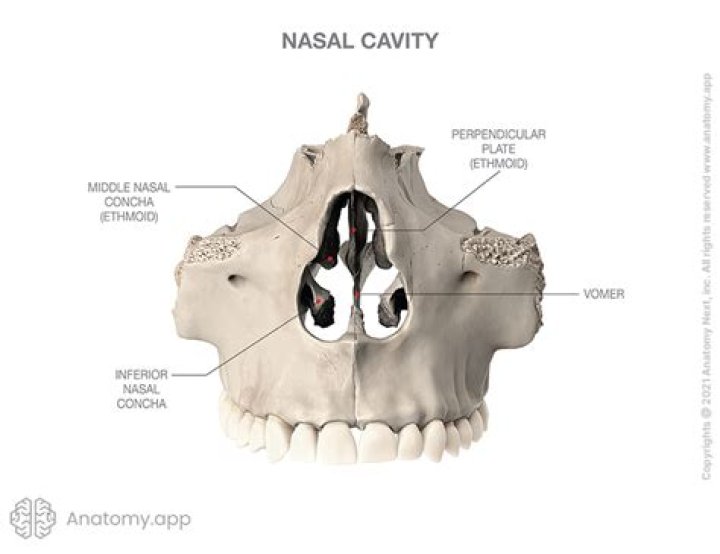
What separates the nasal cavity?The nasal septum divides the cavity into two cavities, also known as fossae. Each cavity is the continuation of one of the two nostrils.
Article first time published onWhat is unilateral choanal atresia?
In unilateral choanal atresia, only one side of the nasal passage is blocked. The baby can still breathe through the side that is not blocked. Choanal atresia. Choanal atresia is a condition in which the back of the nasal passage is blocked by bone or soft tissue. Some babies have a blocked nasal passage on one side.
What is the nasal meatus?
The nasal meatuses are distinct air passages of the lateral nasal cavity located inferior to each nasal conchae.
What is Limen Nasi?
[ nā′zī ] n. A ridge marking the boundary between the nasal cavity proper and the vestibule of the nose.
What are the names of the nasal cartilages?
There are five individual cartilages that make up the nasal cavity: septal nasal cartilage, lateral nasal cartilage, major alar cartilage (greater alar cartilage, or cartilage of the aperture), minor alar cartilage (lesser alar cartilage, sesamoid, or accessory cartilage), and vomeronasal cartilage (Jacobson’s …
What is nasal cavity?
(NAY-zul KA-vuh-tee) The space inside the nose. The nasal cavity lies above the bone that forms the roof of the mouth and curves down at the back to join the throat. It is divided into two sections called nasal passages. Air moves through these passages during breathing.
What is posterior nasal opening called?
Choana is the posterior nasal aperture. The choanae are separated by the vomer.
What is nose bone called?
From Wikipedia, the free encyclopedia. Nasal bone.
Where is the nasal vestibule?
The area just inside the nostril (nose opening) that leads into the nasal cavity. The nasal vestibule is supported by the cartilage of the nose and lined with tissue that contains small, course hairs. These hairs help filter dust, sand, and other particles to keep them from entering the lungs.
What is nasal valve surgery?
Nasal valve surgery is designed to stabilize or support the side walls of the nose. This holds the nasal valve more open during breathing. Nasal valve surgery can be performed by itself or in conjunction with other nasal procedures.
What happens after nasal valve repair?
You can expect to be off work anywhere from a few days to a couple of weeks. Although the incisions made for nasal valve surgery are small and are typically made inside your nose, you will need to keep your head elevated initially, to reduce the chance of bleeding. And you may experience some swelling or bruising.
Does nose block affect breathing?
When your nose feels stuffy, you may find it hard to breathe. The inflammation leads to swollen nasal passages that constrict air flow, making it harder to breathe through your nose. The inflammation and swelling also makes it harder to get mucus out of your nose, so you may also have a build-up of mucus, as well.
What is incisive foramen?
The incisive foramen (also known as nasopalatine foramen or anterior palatine foramen) is the oral opening of the nasopalatine canal. It is located in the maxilla in the incisive fossa, midline in the palate posterior to the central incisors, at the junction of the medial palatine and incisive sutures.
Where is maxillary?
The maxilla is the bone that forms your upper jaw. The right and left halves of the maxilla are irregularly shaped bones that fuse together in the middle of the skull, below the nose, in an area known as the intermaxillary suture.
What is Ostiomeatal complex?
The ostiomeatal complex (OMC) is the collection of structures that aids in mucus drainage and airflow between the maxillary sinus, the anterior ethmoid air cells, and the frontal sinus. It is located on the lateral wall of the nasal cavity and has several well defined borders.
Is nasal cavity connected to brain?
All of the sinuses surround important structures including the brain and eye, so sinus problems can affect both. In fact, your nose is connected to most parts of your head and neck anatomy.
What is meant by concha bullosa?
A concha bullosa is a pneumatized (air-filled) cavity within a nasal concha, also known as a turbinate. Bullosa refers to the air-filled cavity within the turbinate. It is a normal anatomic variant seen in up to half the population.
What is frontal sinusitis?
Frontal sinusitis is inflammation or infection of the sinuses located just behind the eyes and in the forehead. The sinuses are a system of connected hollow cavities in the face that contain air and a thin layer of mucus. All sinuses produce mucus that moisturizes the airways and drains into the nasal passages.
What are 3 main functions of the nasal cavity?
The nasal cavity functions to humidify, warm, filter, and act as a conduit for inspired air, as well as protect the respiratory tract through the use of the mucociliary system. The nasal cavity also houses the receptors responsible for olfaction.
Where does the nasal cavity start and end?
The nasal cavity extends from the external opening, the nostrils, to the pharynx (the upper section of the throat), where it joins the remainder of the respiratory system. It is separated down the middle by the nasal septum, a piece of cartilage which shapes and separates the nostrils.
Which of the followings divide the nasal cavity into right and left halves?
The septum divides the nasal cavity into right and left halves. The nasal cavity is connected with a number of structures. The olfactory nerves penetrate the roof of the nasal cavity and the cribriform plate and innervate the superior nasal concha and the upper third of the septum.
How do you confirm unilateral choanal atresia?
To test for both types of choanal atresia, a medical history review and physical examination will reveal associated symptoms and signs. A test for nasal patency, or airflow is also often performed and a CT scan may be examined to confirm the presence of choanal atresia.